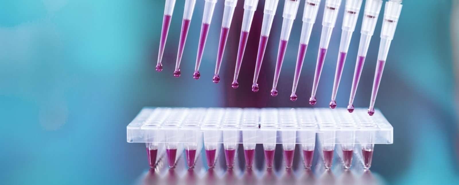
Combining methods.
Obtaining safe results.
We combine a broad range of diagnostic methods to obtain precise findings: To do this we use state-of-the-art devices and technologies that we are able to deploy efficiently thanks to our laboratory concept. Consistent interdisciplinary communication of the various methodical findings is an essential part of our reliable and rapid diagnoses.
During the course of therapy, the detection of measurable residual disease (MRD) becomes increasingly important, which is performed by the highly sensitive methods of molecular genetics and immunophenotyping.
Cytomorphology
The diagnosis of hematological neoplasias is based on cytomorphology and histology. They are used to complete diagnosis, classify diseases and to assess prognoses over the course of treatment. In this regard, cytomorphology mainly involves the assessment of panoptical staining and a variety of cytochemical stains in blood and bone marrow smears. Cytomorphological assessment permits the description and distinction of malignant and healthy cells.
- Bone marrow (Citrate/EDTA)
- Peripheral blood (Citrate/EDTA)
2 hours to 1 day

Chromosome analysis
Chromosome analysis (metaphase cytogenetics) is now considered an obligatory test method for the diagnosis of hematological neoplasia. The findings contribute to substantiating the diagnosis, although the prognostic significance that can be confirmed from the karyotype of malignant cells is far more important. Neoplasia-associated chromosome aberrations occur only in malignant cells. These are acquired genetic mutations. Hence, the other cells in the body of a patient suffering from hematological neoplasia are cytogenetically normal.

FISH
Reflecting the broad variety of chromosome aberrations observed in acute leukemias in particular, FISH “screening” of interphase nuclei only covers a fraction of the abnormalities that may potentially be present and is therefore unable to replace chromosome analysis. However, the FISH technique is a rapid and reliable method that can confirm or exclude a specific genetic abnormality within 4 hours, for instance validation of a translocation t(15;17)(q24;q21) for cases of suspected acute promyelocytic leukemia.

Molecular genetics
Molecular genetics has become an important method in hematology. Thus, it contributes to diagnostics by detecting known genetic aberrations. Through its PCR-based approaches, it shows a high sensitivity, so that remissions, but also relapses can be detected early through quantitative approaches in therapy control. In addition, molecular genetic markers often allow risk stratification or contribute to therapy decisions. Molecular genetic approaches are characterized by the use of DNA or RNA and allow targeted marker detection and increasingly genome-wide, parallelized analyses of different genetic alterations.

Immunophenotyping
Immunophenotyping is key to the diagnostics of hematologic neoplasms. It is used to complete diagnosis, classify diseases, assess prognoses and to quantify malignant cells over the course of treatment (minimal/measurable residual disease, MRD).
Diagnostic application of the method is based on the characterization of antigen expression patterns among malignant cells and their discrimination from healthy cells.

Bioinformatics
Bioinformatics is a relatively new, interdisciplinary field of science that develops and implements methods for the computational analysis, organization, and storage of biological data. The main goal of bioinformatics is to improve biological processes and their pathological changes by developing a deeper understanding of the underlying molecular regulatory networks.

Measurable residual disease (MRD)
The term "measurable residual disease" (MRD) refers to a residual malignant cell population detectable in leukemia patients during or after therapy. Especially in patients with acute leukemias (AML and ALL) and lymphoproliferative diseases, the clinical diagnosis of MRD is becoming increasingly important, as it allows information about the response to therapy as well as the early detection of a relapse.


The increasing diversity of clinically relevant molecular markers is a major challenge for hematological diagnostics. With the introduction of modern high-throughput sequencing methods, also called Next Generation Sequencing (NGS), hundreds of thousands of genome regions can now be analyzed in parallel within a very short time with very high sensitivity.
You may also be interested in
Your contact person

»We achieve higher quality through interdisciplinarity.«
Prof. Dr. med. Wolfgang Kern
Executive Management
Internist, Hematologist and Oncologist
Deputy Head of Immunophenotyping
wolfgang.kern@mll.com