MLL at the 65th ASH Annual Meeting & Exposition - a follow-up report
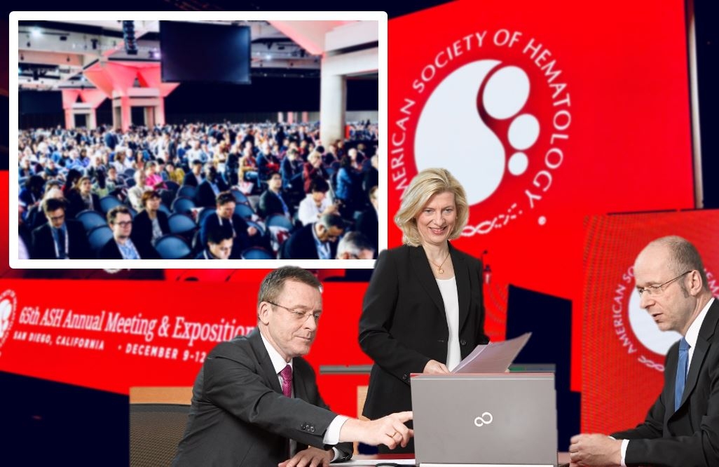
In 2023, the Annual Meeting & Exposition of the American Society of Hematology (ASH) was once again a firm fixture in the calendars of numerous MLL employees. The congress took place for the 65th time, this year from December 9-12 in San Diego, California. MLL was represented with a total of 15 contributions - as a lecture, poster or abstract only.
Artificial intelligence in routine hematology diagnostics
The use of artificial intelligence (AI) in routine haematology diagnostics is a topic that has become an integral part of discussions and symposia. MLL founder and Managing Director Prof. Dr. med Dr. phil. Torsten Haferlach chaired the special interest session "AI in Hematology: Where do you stand in 2023?" at the ASH.
Two other MLL contributions focused on AI applications in immunophenotyping and cytomorphology. Martha-Lena Müller and her colleagues developed an AI model to classify and differentiate mature B-cell neoplasms based on flow cytometry data. Christoph Kornauth and his team investigated the AI-based classification in cytomorphology, whereby the AI was able to identify seven relevant pathological patterns in 872 cases with an acceptable prediction probability.
Focus on a continuum: from CCUS to MDS to AML
The current WHO classification 2022 lists clonal hematopoiesis of undetermined potential (CHIP) and clonal cytopenia of undetermined significance (CCUS) as separate entities and considers them together with myelodysplastic neoplasia (MDS) and acute myeloid leukemia (AML) as a dynamic disease continuum. In her ASH paper, Claudia Haferlach suggested that the classification of CCUS, MDS and AML should be based purely on genetic parameters in order to reflect the biological continuum of these three entities. Reclassification during the course of the disease would thus be possible without changing the entity. Constance Baer has also investigated these entities and presented in a lecture that WHO-defining mutations are mostly early events, which supports their role as defining genetic anomalies. However, other initial mutations indicated that MDS and AML arise from clonal hematopoiesis.
In another presentation, Sandra Huber validated the question of whether peripheral blood can reliably reflect molecular and cytogenetic alterations from bone marrow. A comparison of samples from peripheral blood and bone marrow from 200 patients with cytopenia showed a high degree of overlap, making peripheral blood a reliable surrogate under certain conditions.
In contrast to the WHO, the ICC has introduced an overlapping MDS/AML category, defined by a blast percentage of 10-19% and the absence of recurrent genetic alterations. In a lecture, Gregor Hörmann presented the grouping of an MDS/AML cohort according to the criteria of the European LeukemiaNet (ELN) for risk stratification, but none of the 403 patients met the characteristics for the favorable ELN risk group. For a more adequate risk stratification, the authors (Sandra Huber and colleagues) modified the ELN criteria and thus significantly improved the prognostic stratification.
Further subgroups for AML?
Genetic parameters are playing an increasingly important role in classification and diagnosis. The WHO 2022 now classifies numerous entities into genetically defined subtypes, such as AML. In addition to the existing subtypes, further groupings are suspected. For example, Isolde Summerer postulated myeloid neoplasms with MYC-positive double minutes as a separate, genetically defined entity. The authors base this on their study, in which cases of this subgroup can often be assigned to AML-MR (myelodysplasia-associated), but also show severe dysplasia in the absence of genetic changes that lead to a diagnosis of AML-MR. In addition, these cases show other very characteristic morphological changes, some of which are strongly reminiscent of acute promyelocytic leukemia, as well as similar gene expression profiles and characteristic mutations. Anna Stengel also described a cohort of patients associated with AML who present with the rare cytogenetic alteration i(7)(p10). Angelika Müller-Jochim looked at AML cases with the rare t(4;12)(q12;p13) translocation. The analysis using whole genome and whole transcriptome sequencing allows two subgroups to be distinguished, which are characterized by different breakpoint cluster regions on 4q12 and PDGFRA gene expression.
Further information on research projects at MLL can be found on our website. The links to all MLL presentations at this year's ASH conference can be found here:
Baer C et al. The frequency of clonal hematopoiesis prior to AML and MDS varies among the different molecularly defined WHO subtypes. https://doi.org/10.1182/blood-2023-179737
Ecker V et al. Lymphoma cell sorting to enrich low degree infiltration clonal B-cells for diagnostic delineation of molecular genetic background by NGS. https://doi.org/10.1182/blood-2023-188953
Haferlach C et al. (1) Whole genome and transcriptome sequencing of 21 paired chronic and blast phase CML cases: Acquisition of genomic alterations, changes in the transcriptomic profiles and occurrence of B-cell receptor rearrangements. https://doi.org/10.1182/blood-2023-185867
Haferlach C et al. (2) A proposal for a classification of CCUS, MDS and AML primarily based on genetic abnormalities considering the biological continuum of these entities. https://doi.org/10.1182/blood-2023-179188
Huber S et al. (1) Modification of the ELN classification 2022 refines risk assessment in MDS/AML patients. https://doi.org/10.1182/blood-2023-179822
Huber S et al. (2) Parallel Genomic Analysis from Paired Bone Marrow and Peripheral Blood Samples of 200 Cytopenic Patients. https://doi.org/10.1182/blood-2023-174843
Huber S et al. (3) Genomic Landscape of CCUS Compared to MDS Indicates a Potential Applicability of the IPSS-M. https://doi.org/10.1182/blood-2023-177582
Huber S et al. (4) Classification of Philadelphia-negative MPN as Low Risk and High Risk MPN Based on Peripheral Blood Values and Molecular Genetics Only. https://doi.org/10.1182/blood-2023-174872
Kornauth C et al. Automated Cytomorphological Analysis of Bone Marrow Samples: A proof-of-principle study for AI-based Classification on a Real-Life Data Set of 979 unselected cases. https://doi.org/10.1182/blood-2023-188716
Müller H et al. Can whole genome and whole transcriptome sequencing replace standard procedures in CLL diagnostics? https://doi.org/10.1182/blood-2023-185732
Müller M-L et al. Artificial Intelligence (AI)-predicted medical diagnosis in suspected mature B-cell neoplasms based on flow cytometric raw data. https://doi.org/10.1182/blood-2023-189799
Müller-Jochim A et al. AML with t(4;12)(q12;p13): A detailed genomic and transcriptomic analysis reveals genomic breakpoint heterogeneity, absence of PDGFRA fusion transcripts and presence of PDGFRA overexpression in a subset of cases. https://doi.org/10.1182/blood-2023-180512
Sakuma M et al. M41 and non-M41 UBA1 variants differ in their alteration of hematopoiesis. https://doi.org/10.1182/blood-2023-188640
Stengel A et al. Characterization of Cases with the Rare Cytogenetic Abnormality i(7)(p10) Reveals an Association with IDH2 Mutated Acute Myeloid Leukemia. https://doi.org/10.1182/blood-2023-179844
Summerer I et al. Proposal of Myeloid Neoplasms with MYC-positive Double Minutes as a Distinct Entity. https://doi.org/10.1182/blood-2023-189902

»Sie haben Fragen zum Artikel oder wünschen weitere Informationen zum 65. Meeting der ASH? Schreiben Sie mir gerne eine E-Mail.«
Dr. rer. nat. Constanze Kühn
Medical Writer