Acute lymphoblastic
leukemia (ALL)

Based on the current guidelines and the current state of research, there are different diagnostic recommendations for patients with acute lymphoblastic leukemia. We have summarized the most important information on classification and diagnostic methods at MLL. In addition, we provide further links and literature on prognosis and therapy in ALL.

  • Method:
  • Anticoagulant:
  • Recommendation:
  • Method:
    Cytomorphology
  • Anticoagulant:
    EDTA
  • Recommendation:
    obligatory
  • Method:
    Immunophenotyping
  • Anticoagulant:
    EDTA or Heparin
  • Recommendation:
    obligatory
  • Method:
    Chromosome analysis
  • Anticoagulant:
    Heparin
  • Recommendation:
    obligatory
  • Method:
    FISH
  • Anticoagulant:
    EDTA or Heparin
  • Recommendation:
    obligatory
  • Method:
    Molecular genetics
  • Anticoagulant:
    EDTA
  • Recommendation:
    obligatory

ALL: Classification

According to the WHO classification, ALL is classified together with lymphoblastic lymphoma (LBL) as lymphoid precursor neoplasms of the B- or T-cell type.

The term ALL is conventionally used for cases in which the peripheral blood and bone marrow are primarily affected, and the term LBL for cases in which lymph nodes or extranodal sites are primarily affected. If both the bone marrow and extramedullary organs/sites are affected, the distinction becomes arbitrary - however, many treatment protocols define leukemia when >20-25% blasts are present in the bone marrow. Classification into subgroups (Fig. 1) is primarily based on cytogenetic and molecular genetic criteria (WHO 2022). The immunophenotypic classification according to EGIL, which is used to classify the risk groups of the GMALL study group, is also clinically relevant. In addition, the immunological subtypes are associated with specific clinical and cytogenetic aberrations.
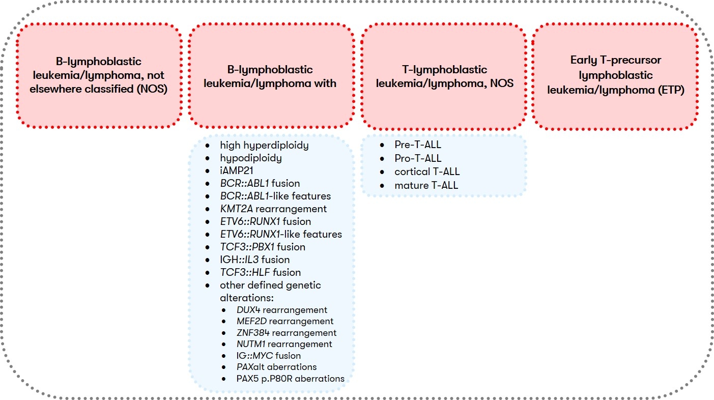
Fig. 1: ALL classification according to WHO (WHO 2022) and GMALL/EGIL

Table 1: Clinical Presentation of ALL (Gökbuget et al., Blood 2024 [2]; Onkopedia 2022, WHO 2022)

Feature

B-ALL

T-ALL

Typical age of onset

At any age, more common in children

More frequent in adolescents and young males

Mediastinal mass

Uncommon

Common (involvement of the thymus)

CNS infiltration

5-10% at diagnosis

Higher risk in T-ALL

Lymphadenopathy

Frequent

Common

Leukostasis symptoms (e.g., shortness of breath, neurological signs)

Possible with hyperleukocytosis

Possible with hyperleukocytosis

For B-ALL (B-lymphoblastic leukemia/lymphoma) and T-ALL (T-lymphoblastic leukemia/lymphoma, NOS), different diagnostic recommendations apply in each case. Therefore, we have summarized them separately for you below:

B-ALL: Diagnostic methods and their relevance

T-ALL: Diagnostic methods and their relevance

ALL: Prognosis and Therapy

The GMALL study divides ALL into standard and high-risk groups based on the following factors: Leukocyte count, immunophenotype, late CR, cyto/molecular genetic aberrations, and MRD. For an overview, see the Onkopedia guideline ALL. The European Leukemia Net (ELN) also provides an overview of therapy management (Gökbuget et al. 2024 [2]).

MRD status during and after therapy influences event-free survival and overall survival (Gökbuget et al. 2024 [2]). MRD is a highly significant prognostic factor and enables early detection of relapse. It is used to rapidly adapt the treatment strategy. MRD can be quantified using molecular genetics or immunophenotyping.

NGS (next-generation sequencing)-based MRD assays show superior sensitivity compared to conventional multiparameter flow cytometry and can detect residual disease in patients who are MRD-negative by flow cytometry. Achieving NGS-based MRD negativity correlates strongly with improved event-free and overall survival and is highly predictive of the risk of relapse after stem cell transplantation and CAR-T cell therapy (Stefan et al. 2025).

The prognostic significance of MRD is influenced by disease-related factors (cytomic molecular risk, immunophenotype) and assay-related parameters (sensitivity, sample material, measurement time). Current expert panels therefore recommend integrating MRD information into clinical decision algorithms for therapy management in the context of these factors (Short et al. 2025).

Due to the complexity and longevity, therapy should be performed in a hematological center.

Biobanking of blood and bone marrow samples is recommended (Gökbuget et al. 2024 [1]) – preferably within a clinical trial or registry. These samples can support future molecular or cytogenetic testing, especially in cases of treatment resistance, relapse, or uncertain therapeutic response. Biobanked material may help identify eligibility for targeted therapies at later stages of treatment.

Table 10: Treatment Response Gökbuget et al., Blood 2024

Feature

B-ALL

T-ALL

Response to standard chemotherapy

Good in standard-risk patients

Similar to B-ALL; subtype-dependent (favorable for cortical T-ALL, poor for ETP/early T-ALL)

Role of stem cell transplantation

Indicated in high-risk cases (poor MRD response, high-risk genetics)

Indicated in high-risk cases (poor MRD response, ETP/immature T-ALL)

Targeted therapy options

e.g., TKIs for BCR::ABL1-positive cases

e.g., NOTCH1 inhibitors , BCL-2 inhibitors (in development)

Status: December 2025

You may also be interested in

Career

As a rapidly growing, innovative medical laboratory, we are always looking for bright minds to help us bring new and more effective therapies to patients around the world.

Learn more

Services

Do you have questions about sample submission, analyses performed or findings? Here you will find contact details, contact persons and our most frequently asked questions (FAQs).

Learn more

Quality management

We have been certified according to national and international standards since 2009 and have successfully maintained these accreditations.

Learn more