Fluorescence in situ
hybridization (FISH)
Use of fluorescent probes for the identification of known chromosome or genetic aberrations in the metaphases or interphase nuclei.
- Bone marrow
- Peripheral blood
1 to 3 days
The FISH technique can be during follow up after therapy as a method for the determination of residual disease whenever the initial diagnosis using chromosome analysis has discovered abnormalities that can be identified by using suitable FISH probes. This method is more sensitive than chromosome analysis, but less so than polymerase chain reaction, next generation sequencing or immunophenotyping.
Test material
Between 5 and 10 ml of bone marrow or peripheral blood anticoagulated with heparin are required for FISH analysis. EDTA or citrate can be used alternatively as anticoagulants. Peripheral blood is sufficient as a test material, provided that malignant cells are present in the peripheral blood. If this is not the case bone marrow should be tested.
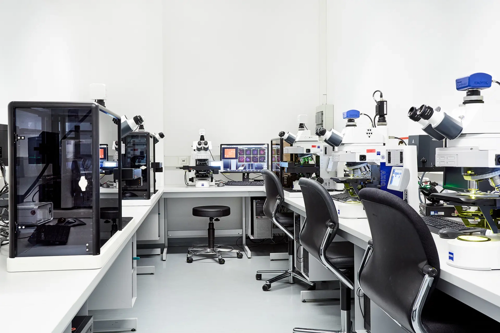
Methodology
Specific hybridization with fluorescence-marked DNA probes
The FISH technique is based on the hybridization of DNA probes that identify specific chromosomal structures. It is possible to use probes that mark specific the centromeric regions of individual chromosomes, genes or entire chromosomes. The DNA of the selected probes and the patient DNA are denaturated, meaning that the two DNA strands of the double helix are separated. During subsequent renaturation, the DNA probes accumulate on the complementary sections of the patient DNA (hybridization). The DNA probes directly carry the fluorescent marker. Therefore, the matching chromosome structures show up as fluorescence signals.
Advantages and limitations of FISH
|
Advantages |
Limitations |
|
Can be conducted on metaphases and interphase nuclei (cell division is not necessary). Identifies even small abnormalities below the resolution limit of chromosome analysis. |
The probes can only provide information about the targeted chromosomes/genes. |
Interphase FISH
FISH can be also conducted on interphase nuclei in addition to chromosome analysis, for screening of abnormalities, e.g. in CLL or multiple myeloma, and during disease progression. The FISH technique is particularly suitable for the identification of so-called sub-microscopic abnormalities below the resolution limit of chromosome analysis, for instance small deletions like those present on 13q in CLL, or tiny insertions that are observed among a small proportion of patients with fusion genes.
The FISH technique can only be used for follow up studies after therapy to determine residual disease if at initial diagnosis chromosome analysis or FISH have identified an abnormality for which suitable FISH probes are available. Interphase FISH is more sensitive than chromosome analysis, but far less so than polymerase chain reaction, next generation sequencing or immunophenotyping.
Metaphase FISH
Besides loci specific probes that can be used on interphase nuclei and metaphases, so-called chromosome painting probes are available that specifically mark the entire DNA of a distinct chromosome. These can only be applied to metaphases. This technique is used to confirm or resolve chromosome abnormalities identified by chromosome analysis in difficult cases. It can also be conducted on the same metaphases analysed after banding analysis.
24-color FISH
The 24-color FISH method identifiesall 22 chromosome pairs, as well as the X and Y chromosome in a single hybridization by depicting them in a different color. It can only be performed on metaphase chromosomes, and is used to resolve complex structural aberrations. It can be conducted on the same metaphases that have been analysed in banding analysis.
Nomenclature
ISCN for FISH on interphase nuclei
According to the ISCN (International System for Human Cytogenetic Nomenclature), Interphase FISH findings begin with “nuc ish” for “nuclear in situ hybridization.” In the formula, the probe is specified by the chromosome band in which it is located and by its precise designation, either as the name of a gene or the designation of an anonymous locus. Furthermore, the formula indicates how many signals for a specific probe were observed and if appropriate its position relative to the other analyzed probes.
Accordingly, in case of a normal finding (2 chromosomes 8 in each cell nucleus) a probe for the centromere region of chromosome 8, will be described by the ISCN formula as “nuc ish 8cen(D8Z2x2),” while in case of a trisomy 8 (3 chromosomes 8 in each nucleus) would be described as “8cen(D8Z2x3).”
If hybridization is performed with probes that cover the breakpoints in genes RUNX1 and RUNX1T1 that are affected by the t(8;21)(q22;q22)/RUNX1-RUNX1T1 rearrangement, the ISCN finding would be, in the case of an RUNX1-RUNX1T1 rearrangement,
nuc ish 8q22(RUNX1T1x3),21q22(RUNX1x3),(RUNX1T1 con RUNX1x2).
The breaks in the RUNX1 gene and the RUNX1T1 gene cause a splitting of the probes and hence 3 signals for RUNX1 and RUNX1T1, respectively: 8q22(RUNX1T1x3),21q22(RUNX1x3); one RUNX1- and one RUNX1T1 signal in each case are located directly adjacent on the two derivative chromosomes (colocalization): (RUNX1T1 con RUNX1x2).
In case of a verified deletion, e.g. in the long arm of chromosome 5, band 5q31, the ISCN interphase FISH finding is “nuc ish 5q31(EGR1x1)” – there is only one signal for EGR1, instead of the usual two.
Downloads
You may also be interested in
Your contact person

»Leukemia diagnostics is becoming more comprehensive as therapy becomes more individualized.«
Prof. Dr. med. Claudia Haferlach
Executive management
Medical doctor
Department manager Diagnostics
claudia.haferlach@mll.com